Background : Hematopoietic stem and progenitor cells (HSPCs) undergo functional changes with age. An unresolved issue is whether aging is induced by a shift in intrinsic state in which all HSPCs undergo coordinated changes in functional potential, or by clonal evolution of a senescent population.Identification of senescent cells in human hematopoiesis poses another challenge. In this study, we have identified lineage-specific, senescence-associated pathways at different developmental stages of HSPCs and have developed an “aging signature HSC” for non-primed human HSCs.
Methods: HSPCs (CD34+ cells) were isolated from the bone marrow of healthy human subjects (old = >60 years; young = 21 to <35 years). Single-cell RNA-sequencing (scRNAseq) studies and downstream analysis were performed using different R packages for normalization, dimension reduction, clustering, classification, pseudotime ordering, and developmental trajectory analyses. Gene Set Enrichment Analysis, GSEA (version 4.3.2., Broad Institute, Inc) was applied to identify pathways and processes that were coordinately up- or down-regulated with aging.
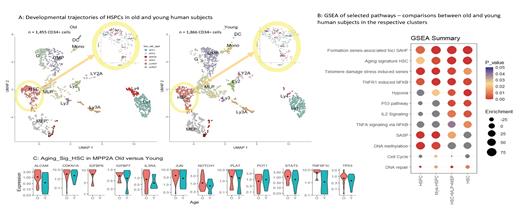
Results: HSPCs (CD34+ cells) were derived from 13 healthy human subjects (old subjects n = 6; young subjects n = 7). There were 26,076 genes identified. The HSPCs were ordered and classified along their developmental trajectory during differentiation processes from (1) hematopoietic stem cells (HSC) to (2) myeloid-lymphoid progenitors (MLP), (3) megakaryocyte-erythrocyte progenitors (MEP), (4) granulocytic-monocytic progenitors (GMP), (5) granulocyte- (G), and (6) monocyte- (Mono) progenitors, (7) dendritic cell (DC), (8) erythroid progenitors (E), as well as various subsets of lymphocytes (Ly1 to Ly4). The results are depicted in Figure 1A. Remarkable are the increases in HSCs (p < 0.05) and in Ly4 (p < 0.001) in old subjects. The Kolmogorov-Smirnov test showed a significant delay in the developmental “pseudotimes” of HSCs (p < 0.001) and of Ly4 (p < 0.05). We then compared the differences in GSEA between old and young subjects in each of the following clusters: (A) whole HSPCs; (B) HSPCs along myeloid pathway (mye-HSPC); (C) HSC-MLP-MEP; (D) HSC. The major findings relevant to aging are summarized in Fig. 1B. Pathways that are significantly increased in the old subjects and throughout all comparisons include: “Formation of senescence associated heterochromatin foci”, “Aging signature HSC”, “DNA damage telomere stress induced senescence”, and “TNFR1 induced NFKB signaling pathway”. “Hypoxia”, “P53 pathway”, “IL2 signaling”, “TNFA signaling via NFKB” were elevated upon enrichment of the primitive HSC population, while other pathways decreased: “Senescence associated secretory phenotype (SASP)”, “DNA methylation”. “Cell cycle” and “DNA repair” are consistently more abundant in young subjects. We analyzed the 512 cells in the HSC cluster with higher resolution and were able to identify a sub-cluster “MPP2A” that is significantly increased in old subjects (4.5% of all HSPCs and 28.5% of HSCs, versus 1.9% and 12.9% respectively in young subjects; p < 0.005). The results are depicted in the highlighted circles in Fig. 1A. This population is quiescent and is highly enriched in “aging signature HSC” in old subjects (p <0.0001). Prominent examples of these signature genes within this list are shown in Fig. 1C). Further along the trajectory, a unique population of B cells “Ly4” was found predominantly in old subjects. The transcriptome profile Ly4 is similar to immature and age-associated B cells (ABCs). This cluster of lymphoid cells may represent the precursors of ABCs described thus far only in peripheral blood, providing another evidence that the aging mechanisms at various differentiation stages are different.
Conclusions: In this study, we have defined specific pathways that distinctly separate aging HSCPs from young ones, and shown that the definition of “aging signature” varies with developmental stages. The accumulation a small cluster in the non-primed HSC compartment, with “aging signature HSC” and cell cycle arrest in old subjects indicates that aging of human hematopoiesis is induced by clonal evolution. The abundance of a unique population among lymphoid precursors with expression profiles similar to ABCs demonstrates that lymphoid and myeloid progenitors age differently.
Disclosures
No relevant conflicts of interest to declare.


